
ဖုဲင်:Epithelial-cells.jpg
resolution တန်ထဲင်းယို အဝ်ႏတဝ်း။
Epithelial-cells.jpg (၂၀၂ × ၂၀၂ pixels, ဖုဲင်ပေႏတန် - ၄၆ KB, MIME အစွိုးအမျိုꩻ: image/jpeg)
| အွဉ်ႏနယ်ချက် |
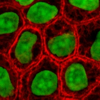
English: Cultured MDCK en:wikipedia:epithelial cells were stained for en:wikipedia:keratin, desmoplakin, and en:wikipedia:DNA. The stained cells were visualized by scanning laser confocal microscopy. The image shows how keratin cytoskeletal filaments are concentrated around the edge of the cells and merge into the desmoplakin which is located at en:wikipedia:desmosomes of the surface membrane. The network of keratin to desmosome to keratin linking the cells of an epithelial sheet is what holds together tissues like skin.
|
||||||||
| ရွီးခိုႏ | wikibooks Cell Biology textbook (licensed under the GFDL): http://wikibooks.org/wiki/Image:Keratin.jpg | ||||||||
| တဲမ်းလိတ်သား | John Schmidt (user:JWSchmidt). | ||||||||
| ဖေႏခွင်ꩻချက် (သုင်ꩻအီချာယင်း ဖုဲင်ယို) |
|

ဖုဲင်မုဲင်တန်ꩻ
ဖေႏကထီႏလꩻ ဖုဲင်နဝ်ꩻမွူးနီꩻအတွိုင်ꩻနဝ်ꩻ နျꩻသွော့ မွူးနီꩻ/အခိန်ႏ တဗာႏဗာႏလောင်း ကလိစ်သြ။
| မွူးနီꩻ/အခိန်ႏ | နမူႏနာႏပွုံႏစံႏပေႏ | ဒုဲင်မဲဉ်းသိဉ်းဖုံႏ | ကေားသုင်ꩻသား | တောင်ꩻသေꩻ | |
|---|---|---|---|---|---|
| ယိုခါကျာꩻ | ၀၂:၁၉၊ ၃ မေ ၂၀၀၅ | | ၂၀၂ × ၂၀၂ (၄၆ KB) | Helix84 | Cultured MDCK epithelial cells were stained for keratin, desmoplakin, and DNA. The stained cells were visualized by scanning laser confocal microscopy. The image shows how keratin [[Cytoskeleton|cytoskele |
သုင်ꩻဖုဲင်
လိတ်မဲ့ငါလ့နဝ်ꩻနဝ်ꩻ အဝ်ႏသွုံꩻခါꩻ ဖုဲင်ယိုသွူ:
ဂလိုဗယ် သုင်ꩻဖုဲင်
ဝီခီအလင်ဖုံႏကို အဝ်ႏသွုံꩻခါꩻ ဖုဲင်ယိုသွူ:
- သုင်ꩻအီ af.wikipedia.org ကို
- သုင်ꩻအီ anp.wikipedia.org ကို
- သုင်ꩻအီ ar.wikipedia.org ကို
- သုင်ꩻအီ az.wikipedia.org ကို
- သုင်ꩻအီ bg.wikipedia.org ကို
- သုင်ꩻအီ br.wikipedia.org ကို
- သုင်ꩻအီ bs.wikipedia.org ကို
- သုင်ꩻအီ ca.wikipedia.org ကို
- သုင်ꩻအီ ca.wikibooks.org ကို
- သုင်ꩻအီ cs.wikipedia.org ကို
- သုင်ꩻအီ cv.wikipedia.org ကို
- သုင်ꩻအီ da.wikipedia.org ကို
- သုင်ꩻအီ el.wikipedia.org ကို
- သုင်ꩻအီ en.wikipedia.org ကို
- User:JWSchmidt
- Tissue engineering
- Intermediate filament
- User:JWSchmidt/Images
- Wikipedia:Facebook directory
- Cytokeratin
- User:Lexor/Temp/Cell (biology)
- Cell culture
- Metabolic engineering
- User:ClockworkSoul/userpage/panel center
- User:ClockworkSoul
- User:Allthesestars/panel centre
- User:SnapJag/userpage/panel center
- Wikipedia talk:WikiProject Molecular Biology/Molecular and Cell Biology/Discussion Archive
- User:Julie Zamostny/BIO134: Cancer Biology
- User:Mesoderm/sandbox
- User:S spoerri/sandbox
- User:Randoperson1/userboxes
- သုင်ꩻအီ en.wikibooks.org ကို
- သုင်ꩻအီ en.wiktionary.org ကို
- သုင်ꩻအီ es.wikipedia.org ကို
နွို့ထွား ကသုင်ꩻခါꩻအီဒါႏဖုဲင်ယို ခင်ႏလမ်းအလင်ဖုံႏကို။